
Main characteristics of patients with Paget‘s disease in a reference hospital
Abstract
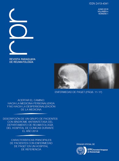
Introduction: Paget‘s disease of bone (PDB) mainly affects adults, with an irregular geographic distribution, with areas of high prevalence, mainly in the United Kingdom, France, and certain regions of Spain. Depending on the radiological findings, the cases are classified in stages: initial or osteolytic; intermediate or mixed and terminal, quiescent or osteosclerotic. The imaging techniques used for the diagnosis are scintigraphy and radiology. Biochemical markers of bone turnover indirectly reflect the activity of PDB and the therapeutic response. The objective of this study is to know the main clinical characteristics of patients with PDB in a reference hospital. Materials and Methods: A retrospective, descriptive, cross-sectional study of patients diag- nosed with PDB at the Vall d‘Hebron Hospital in Barcelona, from January 1985 to December 2009. Data from laboratories, clinics, images and treatment were evaluated. Paget‘s disease of bone was diagnosed by typical X-ray findings. Results: Thirty-five patients diagnosed with PDB were included, with an average age of 61.58 ± 20 years, with an F/M ratio of 1/3. The reason for consultation was pain in 15/35 (42.8%) patients. The most frequent location was hemipelvis, followed by vertebrae, tibia and femur. The involvement in 17/32 (49%) patients was monostotic and in 18/35 (51%) polyostotic. The alkaline phosphatase was elevated between 2 to 10 times the normal value in 30/35 (86%) of the patients at the time of diagnosis. The treatment of choice in all patients was a bisphosphonate, with a response of 57% at 3 months. The most frequent complication was the spinal stenosis. Conclusion: Paget‘s disease of bone affects elderly patients with predominance of the male gender, that may be of polyostotic or monostotic presentation. The most frequent reason for consultation was pain and the most common complication was spinal stenosis. Early diagnosis and treatment with bisphosphonates are very important strategies for these patients.Downloads
References
(1) Barker DJP, Clough PWL, Guyer PB, Gardner MJ. Paget’s disease of bone in 14 British town. Br Med J. 1977;1:1181-3.
(2) Detheridge FM, Guyer PB, Barker DJP. European distribution of Paget’s Disease of Bone. Br Med J. 1982;285:1005-8.
(3) Morales Piga A, Rey Rey J, Corres J, García Sagredo JM, López-Abente G. Frequency and characteristics of the familial aggregation in Paget’s disease of bone. J Bone Miner Res. 1995;10:1-8.
(4) Leach RJ, Singer FR, Roodman G. The genetics of Paget´s disease of the bone. J Clin Endocrinol Metab. 2001;86:24-8.
(5) Murray RO, Jacobson HG. Misellaneous disorders of unknow origin. In.Murray RO. ed. The radiology of skeletal disorders. Edinburg. Churchill Livingtone Eds. 1979: 880-881.
(6) Carbonell J, Rotés D, Maymó J, Lafont A. Principales problemas clínicos en la enfermedad de Paget. Rev Esp Reumatol. 1992;19:104-10.
(7) Gumà M, Rotés D, Holgado S, Monfort J, Olivé A, Carbonell J, et al. Enfermedad ósea de Paget: estudio de 314 pacientes. Med Clin (Barc). 2002;119:537-40.
(8) Schmidek HH. Neurologic and neurosurgical sequale of Paget’s disease of bone. Clin Orthopedic. 1977;127:70-7.
(9) Selby PL, Davie MWJ, Ralston SH, Stone MD. Guidelines on the Management of Paget’s disease of bone. Bone. 2002;31:366-73.
(10) Alvarez L, Guañabens N, Peris P, Vidal S, Ros I, Monegal A, et al. Usefulness of biochemical markers of bone turnover in assessing response to the treatment of Paget’s disease. Bone 2001;29:447-52.
(11) Alexandersen P, Peris P, Guañabens N, Byrjalsen I, Álvarez L, Solberg H, et al. Non-isomerized C-telopeptide fragments are highly sensitive markers for monitoring disease activity and treatment efficacy in Paget’s disease of bone. J Bone Miner Res. 2005;20:588-95.
(12) Farrerons J, Malouf J, Longobardi V, Laiz A. Objetivos e indicaciones del tratamiento farmacológico. Enfermedad ósea de Paget. Barcelona: SCM; 2006. p.91-102.
(13) Ortner DJ. Miscellaneous bone diseases. In: Ortner DJ, editor. Identification of pathological conditions in human skeletal remains, 2nd ed. Amsterdam: Elsevier; 2003.
(14) Vallet M, Ralston SH. Biology and treatment of Paget’s disease of bone. J Cell Biochem. 2016;117(2):289–99.
(15) Mironón-Canelo JA, Del Pino-Montes J, Vicente-Arroyo M, et al. Epidemiological study of Paget’s disease of bone in a zone of the Province of Salamanca (spain). Eur J Epidemiol. 1997;13(7):801–5.
(16) Menéndez-Bueyes LR, Soler Fernández MC. Enfermedad ósea de Paget: aproximación a sus orígenes históricos. Reumatol Clin. 2017;13(2):66–72.
(17) Lisbona Pérez MP, Blanch-Rubió J, Galisteo Lencastre da Veiga C, Esquerra Tuñi E, Monfort Faure J, Ciria Recasens M, et al. Epidemiología de la enfermedad ósea de Paget en un área de Barcelona. Rev Osteoporos Metab Miner 2009 1;1:7-12.
(18) Mautalen C, Pumarino Blanco MC, Gonzalez D. Paget‘s disease: the south american experience. Sem Arthritis Rheum 1994;23:226-7.
(19) Griz L, Caldas G, Bandeira C, Assuncao V, Bandeira F. Paget’s disease of bone. Arqu Bras Endocrinol Metab 2006;50(4):814–22.
(20) Werner de Castro GR1, Heiden GI, Zimmermann AF, Morato EF, Neves FS, Toscano MA, et al. Paget‘s disease of bone: analysis of 134 cases from an island in Southern Brazil: another cluster of Paget‘s disease of bone in South America. Rheumatol Int. 2012 Mar;32(3):627-31.
(21) Falchetti A, Marini F, Masi L, Amedei A, Brandi ML. Genetic aspects of the Paget’s disease of bone: concerns on the introduction of DNA-based tests in the clinical practice. Advantages and disadvantages of its application. Eur J Clin Invest 2010;40(7):655–67.
(22) Favus MJ, Vokes TJ. Paget’s disease and other displasias of bone. In: Longo DL, Fauci AS, Kasper DL, Hauser SL, Jameson JL, Loscalzo J, Eds. Harrison’s Principles of Internal Medicine. New York, NY, USA. McGraw-Hill. 2012. 18th edition. Chapter 355.
(23) Seitz S, Priemel M, Zustin J, Beil FT, Semler J, Minne H, Schinke T, Amling M. Paget’s disease of bone – histologic analysis of 754 patients. J Bone Miner Res. 2009 Jan;24(1):62-9.
(24) Al Nofal AA1, Altayar O, BenKhadra K, Qasim Agha OQ, Asi N, Nabhan M, et al. Bone turnover markers in Paget’s disease of the bone: A Systematic review and meta-analysis. Osteoporos Int 2015;26(7):1875–91.
(25) Muschitz C, Feichtinger X, Haschka J, Kocijan R. Diagnosis and treatment of Paget’s disease of bone. A clinical practice guideline. Wien Med Wochenschr 2017;167:18–24.
(26) Indumathi CK, Dinakar C, Roshan R. Juvenile Paget’s disease. Indian Pediatr 2009;46:354-6.
(27) Ralston SH. Juvenile Paget’s disease, familial expansile osteolysis and other genetic osteolytic disorders. Best Pract Res Clin Rheumatol 2008;22:101-11.
(28) Brunetti G, Marzano F, Colucci S, Ventura A, Cavallo L, Grano M, Faienza MF. Genotype–phenotype correlation in Juvenile Paget disease: role of molecular alterations of theTNFRSF11B gene. Endocrine 2012;42:266–71.
(29) Kumar SR, Bagalad BS, Manohar C, Kuberappa PH. Intermediate type of Juvenile Paget‘s disease: A rare case in Indian population. Contemp Clin Dent 2017;8:175-8.
(30) Wat WZ. Current perspectives on bisphosphonate treatment in Paget’s disease of bone. Ther Clin Risk Manag. 2014;10:977-83.
(31) Papapoulos S, Lippuner K, Roux C, Lin CJ, Kendler DL, Lewiecki EM, et al. The effect of 8 or 5 years of denosumab treatment in postmenopausal women with osteoporosis: results from the FREEDOM Extension study. Osteoporos Int 2015;26(12):2773–83.
(32) Schwarz P, Rasmussen AQ, Kvist TM, Andersen UB, Jørgensen NR. Paget’s disease of the bone after treatment with Denosumab: a case report. Bone 2012;50(5):1023–5.
(33) Reid IR, Sharma S, Kalluru R, Eagleton C. Treatment of Paget’s Disease of Bone with Denosumab: Case Report and Literature Review. Calcif Tissue Int 2016;99:322–5.
(34) Polyzos SA, Cundy T, Mantzoros CS. Juvenile Paget disease. Metabolism. 2018 Mar;80:15-26.
(35) Grasemann C, Schündeln MM, Hövel M, Schweiger B, Bergmann C, Herrmann R, et al. Effects of RANK-lingand antibody (denosumab) treatment on bone turnover markers in a girl with juvenile Paget´s disease. Clin Endocrinol Metab 2013;98(8):3121-6.
Copyright (c) 2018 Paraguayan Journal of Rheumatology

This work is licensed under a Creative Commons Attribution 4.0 International License.